Introduction
Purple urine bag syndrome is a rare phenomenon whose striking presentation can lead to earlier recognition and treatment of a serious medical condition. This is a unique condition of catheter associated urinary tract infections caused by bacterial metabolites. The most important risk factor of developing catheter associated urinary tract infection is length of catherization as risk is 3-10% per day.1 Other risk factors include female sex, diabetes mellitus and bacterial colonization within the urine bag.1 We will demonstrate a case of this rare phenomenon in a patient with squamous cervical cancer who presented to the emergency department with symptoms of a urinary tract infection.
Case Description
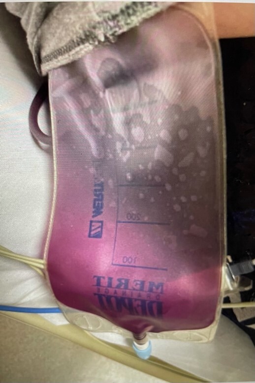
The patient is a 34-year-old G3P3003 with a history of stage IIIb squamous cell cervical cancer diagnosed in December of 2020. Her treatment included 45Gy of external beam radiation with a boost of brachytherapy radiation and only 2 cycles of cisplatin chemotherapy complicated ultimately by loss to follow up. Her cancer course was complicated by bilateral hydronephrosis due to obstruction and subsequent bilateral percutaneous nephrostomy tube (PCN) placement. She presented to the emergency department with decreased urinary output from the bilateral PCNs and left flank pain. She reported decreased urinary output from the PCNs over the preceding month. She expressed she was supposed to have the nephrostomy tubes removed four to five weeks after the second exchange; however, they have remained in place for seven months. The patient also accounted she last changed the nephrostomy bags four weeks prior to her presentation to the emergency department. She mentioned that she was voiding via urethra but also noticed increased urinary frequency and urgency as well as accompanied persistent back pain. She denied dysuria or hematuria. Of note, the patient reported having change in urine color in the nephrostomy bags. She remarked that two weeks prior to her presentation to the emergency department, the color started to change from a yellow-green hue before transitioning to a purple color. She had experienced intermittent chills, but she denied having a fever. The patient was noted to be mildly hypertensive, non-tachycardic and afebrile while in the emergency department. Her physical exam was significant for left costovertebral tenderness and bilateral PCN bags with purple urine. There was no evidence of leukocytosis or electrolyte abnormalities on laboratory values. A urine analysis was collected and significant for pyuria and proteinuria. A urine culture obtained was noteworthy for >100,000cfu/ml proteus mirabalis. Blood cultures were drawn and found to have no growth to date for more than five days. The patient received intravenous antibiotics and intravenous fluids to treat urine tract infection in the emergency room. She was admitted to the hospital but unfortunately left against medical advice shortly after admission. She re-presented to an outside hospital with fevers and non-draining nephrostomy tubes associated with purulent discharge at the nephrostomy sites. The scant urine through the nephrostomy tubes was also noted to have a purple hue. She was found to have another urinary tract infection with Enterobacter cloacae treated initially with intravenous antibiotics. During this admission, she underwent bilateral nephrostograms notable for significant clogging of tubes and bilateral ureteral patency. After the study, she subsequently had her PCNs removed. She was discharged in good and improved condition on a ten-day course of outpatient oral antibiotics.

Discussion
Evaluation of patient urine is not a new tool for assessment of disease. There is data to suggest that ancient civilizations performed urinalysis and uroscopy. “Honey urine” has been described in ancient Sanskrit literature as sweet to the taste with the ability to attract black ants, later determined to be secondary to glucosuria as seen in diabetes.2 In the 14th century, with the aid of matula and urine charts, it was believed that one could diagnose several diseases based on urine color alone.2 From urine charts, taste testing, and now modern-day urinalysis, assessment of urine has become one of the mainstays of initial patient evaluation.
Purple urine bag syndrome was first described in 1978 when it was noted that urine in collecting bags turned purple. After spectral analysis of the urine, it was observed that indigo was present. At that time, it was hypothesized that increased bacterial metabolism in the intestinal lumen resulted in indoxyl sulfate. This would be absorbed and excreted which oxidized to indigo upon contact with the air in the collection bag, resulting in purple colored urine.3 It is also postulated that King George III may have also had purple urine after constipation in the setting of presumed acute intermittent porphyria. This constipation would lead to increased dietary tryptophan metabolism, subsequent indole production, metabolism to indoxyl sulfate, and later indigo blue formation.4
In recent years, purple urine in bag syndrome (PUBS) has been defined as a condition characterized by urine with purple discoloration in patients with urinary tract infection. As detailed in Table 1, other risk factors include indwelling catheterization, female sex, elderly age, asymptomatic bacteriuria, chronic kidney disease, urine alkalinization, and constipation. Conditions that lead to dietary tryptophan having increased time spent in the digestive tract have been considered predisposing conditions for PUBS (constipation, malabsorption, etc).5–8
The pathophysiology behind this condition has further been explained by digestion of dietary tryptophan by intestinal bacteria to indole that is then absorbed into the circulation. Later, in the liver, it is acted on by cytochrome P450 2E1 (CYP2E1) and sulfotransferase 1A1 (SULT1A1) isoform to become indoxyl sulfate. This substrate returns to circulation and is excreted in the urine where in the presence of bacteria with sulfatase and phosphatase activity converts indoxyl sulfate to indigo and indirubin resulting in the characteristic hue of the urine. Providencia stuartii, Pseudomonas aeruginosa, Escherichia coli (E.coli), Proteus mirabilis, Morganella morganii (M. morganii), Klebsiella pneumoniae, Providencia rettgeri, Citrobacter species, Enterococci, and Group B Streptococci are the most common bacteria with the sulfatase and phosphatase activity required.5–10
PUBS is generally considered a benign process; however, treatment should not be delayed as overall mortality related to PUBS has been reported as 6.8%.11 If the infection is not treated it can progress to sepsis. Prior literature reviews have identified rare cases where PUBS progressed to Fournier’s gangrene or multi-drug resistant vulvar abscesses.5,10–14 Indoxyl sulfate has been noted to accumulate in the serum in patients with chronic kidney disease. In the serum it is primarily protein bound and is not readily cleared during dialysis since only unbound indoxyl sulfate can diffuse through the membrane. Accumulation of indoxyl sulfate has been implicated in contributing to further renal failure, vascular disease, cognitive impairment.1 However, the majority of these effects have primarily been assessed in vitro or in animal models. Human toxicity will need further research. When PUBS is suspected further medical evaluation and treatment should be sought as soon as possible.
Once identified, treatment of the urinary tract infection should be aimed at causative bacteria. Additionally, any predisposing condition (constipation) or risk factors should be addressed. This often entails intravenous antibiotics until the urine has returned to normal coloration followed by transition to oral antibiotics, exchange of the urinary catheter, bowel regimen, or evaluation of renal status in CKD patients.5,6,10–13 If a patient presents sepsis or shock, treatment for underlying infection and resuscitation should be initiated. Based on literature review, patients tend to have resolution of purple discoloration with completion of antibiotic treatment and exchange of urine bag.15,16 As with the patient, she initially had initiation of empiric antibiotic treatment; however, left against medical advice prior to completion of medical management. She had a delayed course in care but ultimately completed treatment with directed antibiotics for her urinary tract infection and with removal of percutaneous nephrostomy tubes. Reports of progression of disease in patients who initially presented with purple urine and asymptomatic bacteriuria support the need for treatment and further evaluation in a patient with sudden development of purple urine who is otherwise asymptomatic.1
Conclusion
Health care providers need to be aware of purple urine bag syndrome as it is a condition that can often be diagnosed with visual evaluation of a patient’s urine. Although in many cases benign, without treatment, urinary tract infections can lead to pyelonephritis, sepsis, bacteremia and even death.17 The best prevention of catheter associated urinary infections is stewardship of indwelling catheters including sterile placement and prompt removal as soon as catheter is no longer needed.
Conflict of Interest
There is no conflict of interest to report.
Disclosures
There are no disclosures. Patient consent was obtained.
Author Contribution
Rhea Eubanks, MD: Conceptualization, Methodology, Writing- Original Draft, Writing- Review and Editing Crystal Dupont, MD: Methodology, Writing-Original Draft, Diane Fru, MD: Methodology, Writing-Original Draft Amanda Jackson: Writing- Review and Editing Thomas Herzog, MD: Writing-Review and Editing Caroline Billingsley, MD: Writing-Review and Editing, Supervision