This website uses cookies

We use cookies to enhance your experience and support COUNTER Metrics for transparent reporting of readership statistics. Cookie data is not sold to third parties or used for marketing purposes.

Skip to main content
null
NAPGO
  • Menu
  • Articles
    • Case Report
    • Conference Abstracts
    • Conference Bulletin
    • Expert Reviews
    • Original Research
    • Systemic Reviews
    • All
  • For Authors
  • Editorial Board
  • About
  • Issues
  • search
  • X (formerly Twitter) (opens in a new tab)
  • RSS feed (opens a modal with a link to feed)

RSS Feed

Enter the URL below into your favorite RSS reader.

http://localhost:21113/feed
ISSN 2769-5336
Case Report
Vol. 3, Issue 2, 2024April 05, 2024 EDT

Case Report: Dichorionic-Diamniotic Twin Pregnancy Complicated by a Migrational Anomaly Mimicking a Brain Neoplasm

Eric Schmitt, MD, Pablo Delis, MD, Sonia Pinto, M.B.B.S, Veronica Rooks, MD, Donald Gloeb, DO, MPH,
AnencephalyBrain NeoplasmFetal Gestational AnomaliesCerebral AnomalyMultifetal GestationDichorionic-Diamniotic Twin PregnancyFetal Brain Migrational AnomalyFetal Brain TumorFetal MRIFetal Neoplasm
Copyright Logoccby-4.0 • https://doi.org/10.54053/001c.115908
Photo by National Cancer Institute on Unsplash

Articles in Vol. 3, Issue 2, 2024

Vol. 3, Issue 2, 2024
  • Wellness and diversity, equity, and inclusion support across residencies in core specialties - a national cross-sectional study
    Maggie WongLena FaiselDani ZoorobMalik MaysDanielle DeverJames Van Hook
  • Virtual schooling during the covid pandemic: the effect on high school retention for adolescent mothers
    Beatrice SoderholmJara CrawfordTracey WilkinsonDavid Haas
  • Vaginal cuff dehiscence with small bowel prolapse 10 years after vaginal hysterectomy
    Brandon MorehartDennis SchumacherDennis Lutz
  • Utilization of enhanced recovery after surgery (eras) protocol in gynecologic surgery
    Rachel HartmanSanela AndelijaNatalie HurlockJames BaronStephen ZweibachBradley SipeTiffany TonismaeMiriah Denbo
  • Uterine rupture secondary to pyomyoma leading to intraabdominal abscesses following an uncomplicated vaginal delivery
    Olga Colón-MercadoRachel HartmanJames BaronStephen ZweibachBradley SipeTiffany TonismaeLauren Davis
  • Uterine didelphys with transverse vaginal septum: a case report and review of the literature
    E. Baylee EdwardsKatherine GretteNicolette HollidayCandice Holliday
  • Twin-twin transfusion syndrome in the setting of covid-19 pneumonia: a case report
    Callie HamaiMegan WhiteTuhlei BreannaEric JohannesenElla SpeichingerAlbert Hsu
  • The impact of covid-19 vaccination on intrauterine fetal demise rates among pregnant women in the Metro-Detroit area
    Nicolina SmithMary CondonMariam AyyashGregory Goyert
  • The face of wellness, diversity, equity & inclusion: a national review of obstetrics & gynecology programs
    Malik MaysDani ZoorobLena FaiselDanielle DeverShahnaz VellaniJames Van Hook
  • The current state of medical informatics curricula in undergraduate medical education: a national survey
    Lauren LeibyAnita TamirisaDani ZoorobCatherine VanHook
  • Supplementing provider counseling with an educational video for scheduled induction of labor
    Kelsey PapeSierra CroweDavid Haas
  • Sorrows and the joys of tomorrow: analyzing maternal events during delivery and post-partum in the covid-19 pandemic through utilizing the epds
    Ala Addin Sid AhmedMaheen IrshadFederico Mariona
  • Second case of spontaneous left ureter rupture during normal spontaneous vaginal delivery
    Emily DeckerCollette LessardDennis Lutz
  • Sacral neuromodulation in a patient with pseudocholin-esterase deficiency
    Nadia MegahedPrapti SinghYaseen MalekiJames KirbyJulie Stewart
  • Religious and cultural diversity support across US based obstetrics and gynecology residency program
    Nabila AzeemDani ZoorobYasmin HasbiniJuliana ColvinAriel CohenJames Van Hook
  • Reduction in birth rates and preterm births following the emergence of COVID-19 pandemic in Michigan
    Mariam AyyashMegan McNittGregory Goyert,Madhurima KeerthyMonique Swain
  • Racial bias in cesarean decision making
    Sarah EdwardsQuetzal ClassCatherine FordTamika AlexanderJonah Fleisher
  • Race/ethnicity bias and its role in severe maternal morbidity: a clinical perspective
    Elliot LevineLeah DelfinadoCarlos Fernandez
  • Pregnancy outcome after placement of laminaria and subsequent second trimester removal
    Margaret MonroeTiffany TonismaeRaphael Guichard
  • Preferences and satisfaction of pregnant patients and ob nurses regarding monoclonal antibody infusions for the treatment of covid-19 pneumonia
    Katherine ChenDani ZoorobDavid HarperTracy BuderSamantha AchaeurJames Van Hook
  • Perinatal and maternal outcomes in singleton preterm births of african american versus caucasian women with hypertensive disorders
    Chinemelum OrizuJose PrietoTiffany Tonismae
  • Patient variables which lead to election of early vs. late post cesarean section discharge
    Benjamin KitkoMelissa VassasJames WilsonMichael Krew
  • Partner support has limited influence on clinical outcomes in pregnant patients with substance use disorder
    Neil PatelCynthia CockerhamLeon SuArnold StrombergBarbara ParillaJohn O'Brien
  • Optimal glucose testing for the detection and diagnosis of prediabetes and type ii diabetes mellitus in reproductive age women with polycystic ovarian syndrome
    Wenqi OuyangJaycee HoushRaquel IbarraKathleen GroeschTeresa WilsonPaula Diaz-SylvesterKristin DelfinoJ Ricardo Loret de Mola
  • Obstetrical and neonatal outcomes of pregnancies complicated by sarcoidosis
    Erin EganLaura DiGiovanniChristian AscoliNadera Sweiss
  • National chorioamnionitis trends: Geospatial analysis of vulnerable communities
    Joanne ChanNicholas PetermanBradley KapturRachel MannBrittany VillKristine Carpenter
  • Maternal and infant morbidity and mortality in extremely premature infants - a single centered study in detroit michigan between 23-26 weeks of gestation
    Mariam AyyashKrishna MajmundarGayathri VadlamudiDavid DaCostaGregory Goyert
  • Low-dose aspirin initiation rates in appropriate candidates at an academic residency program in the midwest
    Alexandra Mahdasian-MillerShachia JacksonDavid HaasCaroline Rouse
  • Impact of previous gynecologic care and gynecology service consults on abnormal uterine bleeding follow-up
    Khloe PastoreCaley PolkinghornNicolette CodispotiAnne TjadenLinda Yang
  • Identifying the palliative care needs of patients and families in gynecologic surgical oncology
    Klaudia KukulkaKarle WashingtonMary FaurotRachel AlexanderKevin CraigJean GoodmanErin Tuller
  • Hot yoga for pregnancy: a survey of hot yoga studio owners
    Elizabeth NikcevichViann Nguyen-FengBethany HansonShilpa Babbar
  • Homeopathic treatments of vulvovaginal candidiasis on youtube
    Breia ReedMaya BraggVeronica Mallett
  • Go nuts! Coconut oil as a patient-preferred alternative to commercial ultrasound gel
    Claire EdelmanCaroline RouseZiyi YangJoanne DaggyAnthony Shanks
  • Gastroschisis: To cut or not to cut?
    Mariam AboulelaAntonio Santos RocaAditi DeyPaul DanielsonTiffany Tonismae
  • Female dysorgasmia: an analysis of 19 patients who experience pain with orgasm
    Breton BarrierRaymond FosterMolly Greenwade
  • Factors that influence the choice for neonatal resuscitation in periviable deliveries
    Jessica YoungDiane CasiniTodd FosterTiffany TonismaePeter MarcusKelsey Montgomery
  • Evaluation of socio-economic and immune factors related to endometrial cancer
    Aneesh ChawlaStacy ParrTaylor BadgerEmily DirksKathleen GroeschTeresa WilsonPaula Diaz-SylvesterKristin DelfinoSarah NelsonJ. Ricardo Loret de MolaLaurent BrardAndrea Braundmeier-Fleming
  • Equitable time to treatment of severe hypertension among races at Cleveland Clinic Akron General
    Sarah DavisEstee GeorgeDavid BiatsRachelle AbdelnourCaroline MangiraNatalie Bowersox
  • Effect of pre-operative counseling on post-operative outcomes at the time of minor gynecologic surgery: a randomized control study
    Priscilla TingJason HoppeMichael Krew
  • Deciphering maternal ethnicity from state birth certificates: a perplexing quandary
    Jessica NelsonTiffany AzzarelloSteffen ChristensenThomas ArnoldDennis Lutz
  • Comparing delivery outcomes of concurrent use of mechanical dilation with low dose versus medium dose pitocin versus balloon catheter only
    Victor FeldbaumMaurice HordTiffany Tonismae
  • Colorectal adenocarcinoma presenting as a bowel obstruction in the second trimester of pregnancy
    Thomas ArnoldTracer OverlandDennis Lutz
  • Case of thoracoamniotic shunt placement for treatment of pleural effusion in the setting of congenital diaphragmatic hernia complicated by hydrops
    Antonio Santos RocaKelsey MontgomeryTiffany TonismaeDavid Kays
  • Buprenorphine induction using microdosing for the management of opioid use disorder in pregnancy
    Barbara Parillia
  • Atypical presentations of dicer1 syndrome in adults with juvenile granulosa cell tumor
    Callie HamaiSamantha SalmonArwa MohammadJean GoodmanMark Hunter
  • Atypical cerclage indication: a case report and review of the literature
    Rennan ZahariasWilliam PerezNicolette HollidayCandice Holliday
  • Association of gabapentin concentrations with opioid use after cesarean delivery
    David HaasRebecca SilvolaBobbie RayShelley DowdenAndrea MastersSara Quinney
  • Association between pregnant patients' characteristics and obtaining a biologic drug test
    Monika StoskuteAlexis BravermanDhanalakshmi Thiyagarajan
  • Arab american outcomes of pregnancies complicated by fetal growth restriction
    Nicolina SmithMichelle MillerRobyn GarciaGregory GoyertD'Angela Pitts
  • Application of the ovarian-adnexal radiological data-reporting system (o-rads) for ultrasound diagnosis and management of adnexal masses
    Carlos FernandezElliot LevineAbraham ShashouaIrma SodiniJacqueline LunaPena Maria de la Luz
  • A case series of four women with clear cell ovarian cancer, with unexpected atypical endometriosis as precursor lesions
    Abby LombardiEric JohannesenJane McElroyMark HunterErin TullerAlbert HsuJean Goodman
  • A Low Budget Perineal Repair Model
    Eric SchmittJose RobleJamie Humes
  • Conference Bulletin: Central Association of Obstetricians and Gynecologists - 2022
    North American Proceedings in Gynecology and Obstetrics
  • Donor Screening: Level of Sensitivity in Karyotype with GTG banding vs PGT-A with NGS
    Naveena DaramMeghan Ozcan
  • Contained Uterine Rupture Managed Conservatively after Vaginal Delivery
    Eric SchmittThomas ChoiJose RobleJohmark FarlettAbigail Schmolze
  • A systematic review of maternal exposures and the risk of autism spectrum disorders in their infants
    Shae N JansenKyle M BaughJames B AdamsChristina K FlynnDavid M Haas
  • Case Report: Dichorionic-Diamniotic Twin Pregnancy Complicated by a Migrational Anomaly Mimicking a Brain Neoplasm
    Eric SchmittPablo DelisSonia PintoVeronica RooksDonald Gloeb
NAPGO
Schmitt, Eric, Pablo Delis, Sonia Pinto, Veronica Rooks, and Donald Gloeb. 2024. “Case Report: Dichorionic-Diamniotic Twin Pregnancy Complicated by a Migrational  Anomaly Mimicking a Brain Neoplasm.” North American Proceedings in Gynecology & Obstetrics 3 (2). https://doi.org/10.54053/001c.115908.
Save article as...▾
Download all (7)
  • Figure 1. Abdominal ultrasound, GA 16w1d, Baby A transverse cephalic view.
    Download
  • Figure 2. Twin A in a 24-year-old gravida 1 with a dichorionic-diamniotic twin pregnancy.
    Download
  • Figure 3. Abdominal ultrasound, GA 20w0d, Baby A transverse cephalic view.
    Download
  • Figure 4. A second fetal MRI at 23w0d demonstrated a supratentorial left hemispheric mass within the cerebrum of Twin A.
    Download
  • Figure 5. An axial T1 FLAIR image showing polymicrogyria in the right inferior frontal lobe.
    Download
  • Figure 6. MRI images from day-of-life 1
    Download
  • Figure 7. Post contrast Coronal T1 images showed no enhancement of suspected tumor.
    Download

Error

Sorry, something went wrong. Please try again.

If this problem reoccurs, please contact Scholastica Support

Error message:

undefined

View more stats

Abstract

Background

Fetal brain anomalies have a wide differential diagnosis including: (1) congenital genetic and vascular disorders; (2) acquired causes such as infection and hypoxic-ischemic injury; and rarely (3) neoplasm. Fetal brain anomalies are also infrequently seen. Consequently, there is limited literature describing methods of distinguishing these conditions, barring the presence of the most characteristic findings on imaging.

Objective

The purpose of this report is to describe the critical parts of our workup that helped us distinguish a fetal brain migrational disorder from a neoplasm.

Study Design

Case report of a single patient seen at Tripler Army Medical Center and Children’s Hospital of Los Angeles

Case Summary (Results)

24-year-old gravida 1 with a dichorionic-diamniotic twin pregnancy was found to have an intracranial abnormality in Twin A on a transabdominal ultrasound performed at 16w1d gestation. Serial prenatal ultrasounds and complementary fetal magnetic resonance imaging (MRI) studies were performed to distinguish a possible fetal brain tumor from a migrational disorder. Fetal MRIs performed at 19w1d and 23w0d demonstrated a mass-like region increasing in size which was interpreted as highly suggestive of an enlarging fetal brain tumor. Ultimately, additional radiologic findings of fluid filled clefts and polymicrogyria visualized at 32 weeks led to a definitive diagnosis of a fetal migrational disorder.

Conclusion

Features used to diagnose fetal brain anomalies are often nonspecific and develop throughout a gestation. Most of these diagnoses require multiple imaging studies carried out over time to identify key features, and characteristic key features often do not present until very late in a gestation. The differential diagnosis of a dysplastic appearing mass-like region growing in size throughout a pregnancy should include fetal migrational disorders.

Fetal brain anomalies have a wide differential diagnosis including: (1) congenital genetic and vascular disorders; (2) acquired causes such as infection and hypoxic-ischemic injury; and rarely (3) neoplasm (Jansen and Keymolen 2019). Fetal brain anomalies are seen infrequently. Consequently, there is limited literature describing methods of distinguishing these conditions, barring the presence of the most characteristic findings on imaging.

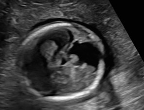
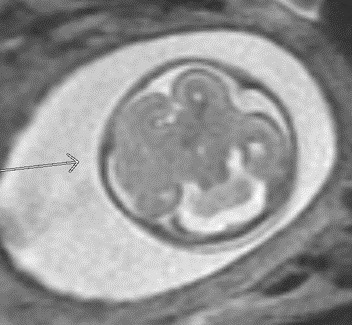
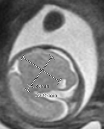
A 24-year-old gravida 1 with a dichorionic-diamniotic twin pregnancy at 16w1d gestation was evaluated by Maternal-Fetal Medicine who identified multiple possible cranio-facial abnormalities in Twin A on transabdominal ultrasound. This raised the concern for a possible chromosomal abnormality as well as holoprosencephaly as the cavum septum pellucidum was not visualized. A detailed transabdominal ultrasound performed at 16w1d provided the initial assessment of the abnormal-appearing left cerebrum of Twin A (Figure 1). The left cerebral ventricle was overall poorly visualized, however, the posterior aspect of the left ventricle appeared to be significantly more dilated compared to the normal-appearing right ventricle. The cavum septum pellucidum could not be visualized, the thalami appeared fused, and the temporal areas of the facies appeared more angulated than expected. The patient was scheduled for a fetal MRI which was performed at 19w1d gestation and showed complete architectural distortion involving the left cerebral hemisphere of Twin A, with a mass-like structure measuring approximately 2.4 x 2.3 x 1.8 cm, along with a loss of normal gray-white differentiation (Figure 2). The diagnoses based on this MRI were either a neoplastic growth or a migrational anomaly.
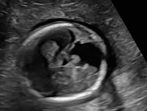
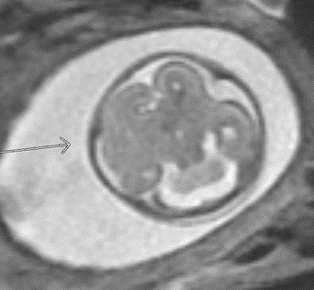
Figure 1
Figure 1.Abdominal ultrasound, GA 16w1d, Baby A transverse cephalic view.

The left cerebral ventricle was overall poorly visualized, however, the posterior aspect of the left ventricle appeared to be significantly more dilated compared to the normal-appearing right ventricle. The cavum septum pellucidum could not be visualized, the thalami appeared fused, and the temporal areas of the facies appeared more angulated than expected.

Figure 2
Figure 2.Twin A in a 24-year-old gravida 1 with a dichorionic-diamniotic twin pregnancy.

A fetal MRI at 19w1d showing complete architectural distortion of the left cerebral hemisphere with a mass-like structure measuring 2.4 x 2.3 x 1.8 cm along with a loss of normal gray-white differentiation.

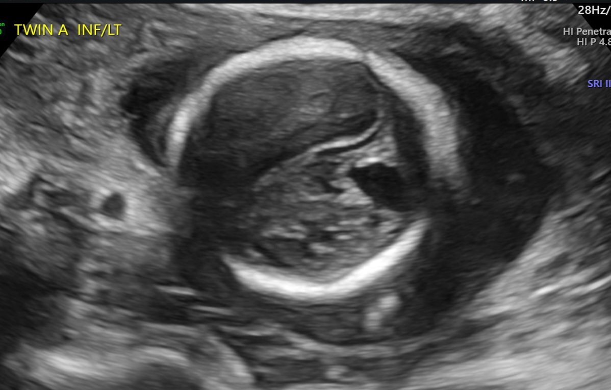
At 20w0d gestation, the patient returned for a repeat ultrasound study which showed a left cerebral mass with a dysplastic appearance, and evident mass effect on the ipsilateral ventricle (Figure 3). The expanded differential at that time included a fetal brain mass, with the highest suspicion for a teratoma. Less likely etiologies included a malformation secondary to a cerebral vascular accident and a congenital brain malformation. As no in utero interventions were indicated based on the suspected diagnosis, focus was directed toward confirming the presence of a fetal brain tumor. Relative growth of this suspected mass over time, measured by a repeat fetal MRI, was possibly suggestive of an aggressive fetal neoplasm.

Figure 3
Figure 3.Abdominal ultrasound, GA 20w0d, Baby A transverse cephalic view.

Images showed a left cerebral mass with a dysplastic appearance, and evident mass effect on the ipsilateral ventricle

At 23w0d a limited fetal MRI was repeated (Figure 4). This study demonstrated, again, the supratentorial left hemispheric mass measuring 3.3 x 3.5 x 2.4 cm within the cerebrum of Twin A. Most notably, there was an interval increase in the size of the mass relative to the skull diameter, as well as a prominent mass effect on the contralateral hemisphere with an associated midline shift. This second fetal MRI finding was interpreted as most consistent with an enlarging intracranial neoplasm with the differential diagnoses including astrocytoma, intracranial teratoma, and glioblastoma multiforme; however, no distinctive features were noted to confirm one of these diagnoses. The interval growth of the mass was interpreted as characteristic of an aggressive fetal neoplasm; therefore, a migrational anomaly was seen as less likely.
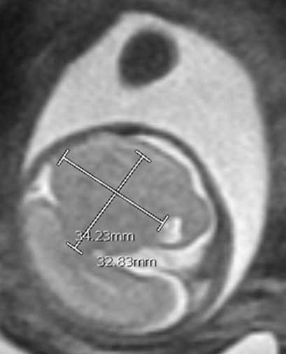
Figure 4
Figure 4.A second fetal MRI at 23w0d demonstrated a supratentorial left hemispheric mass within the cerebrum of Twin A.

This mass had increased in size to 3.3 x 2.5 x 2.4 cm with an associated mass effect on the ipsilateral hemispheric structure and a mass effect on the contralateral hemisphere with a midline shift.

Anticipating the scope of Twin A’s neonatal care needs, it was recommended that the patient deliver at a center with neuro-oncology services and a robust neonatal support center. The patient was transferred to Children’s Hospital Los Angeles for further care. At 30w5d, a prenatal ultrasound study revealed an echogenic nonvascular intracranial mass in the left hemisphere measuring 4.18 x 3.57 cm with significant mass effect along with ventricular dilation. A repeat fetal MRI at 32w0d gestation confirmed a markedly abnormal mass-like configuration in the left cerebral hemisphere and absence of a cava septum pellucidum, basal ganglia, corpus callosum, and right ventricle. Additionally, this MRI had findings suggestive of extensive migrational abnormalities throughout the left cerebral hemisphere, best seen superiorly. It noted no solid or cystic masses and no areas of abnormally restricted diffusion or hemorrhage. While a congenital brain tumor could not be ruled-out, this MRI study reordered the differential diagnosis that had previously favored fetal brain neoplasm.

The patient continued her obstetric care at the Children’s Hospital Los Angeles, and at 35w3d, presented with preterm prelabor rupture of membranes. Both infants were delivered via cesarean section without complication.

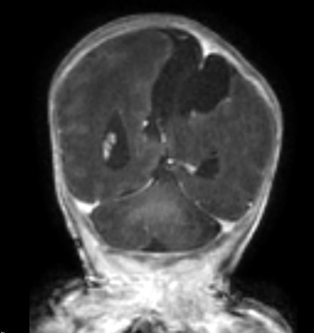
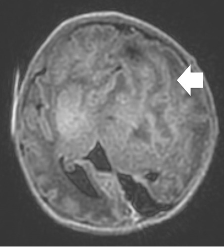
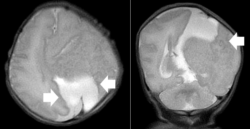
A repeat MRI on day-of-life 1 revealed multiple intracranial anomalies including extensive polymicrogyria of the left cerebral hemisphere, polymicrogyria in the right frontal lobe, incomplete development of the left lateral ventricle, and a gray matter lined cerebrospinal fluid cleft in the left posterior-frontal and parietal regions communicating with the rudimentary left lateral ventricle (Figures 5-7). The dysplastic left cerebral hemisphere exerted a mass effect on the rudimentary left lateral ventricle with a left-to-right midline shift measuring approximately 6 mm at the level of the foramen of Monroe. There was evidence of partial fusion of the frontal lobes. In summary, the first postnatal MRI confirmed that rather than a growing neoplasm, Twin A had extensive intracranial developmental anomalies, including a dysplastic left cerebral cortex, schizencephaly, and hypoplasia or absence of multiple midline structures.

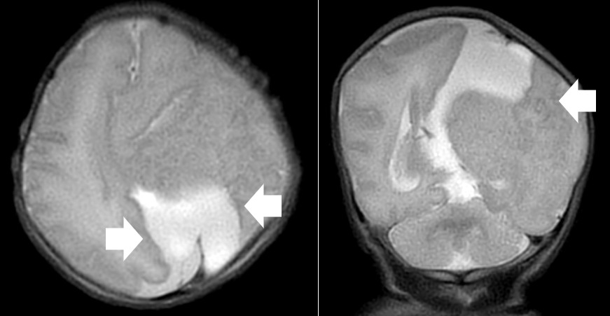
Figure 5
Figure 5.An axial T1 FLAIR image showing polymicrogyria in the right inferior frontal lobe.
Figure 6
Figure 6.MRI images from day-of-life 1

On left, is an axial T2 view showing a schizencephalic cleft in the left parietal lobe along with polymicrogyria. On the right is a coronal T2 view also showing a schizencephalic cleft with polymicrogyria most notably in the adjacent left parietal lobe (long arrow).

Figure 7
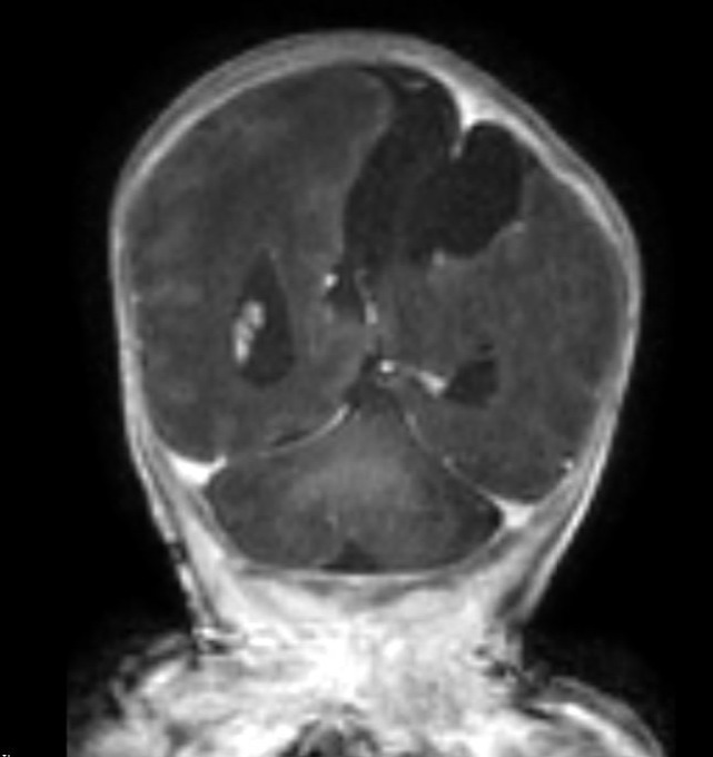
Figure 7.Post contrast Coronal T1 images showed no enhancement of suspected tumor.

The appearance of the malformation on pre-contrast T1 and T2 images and the absence of contrast enhancement after addition of gadolinium is diagnostic of a cerebral malformation and excludes tumor.

Twin A received NICU care at Children’s Hospital of Los Angeles until 5w5d of life, when the infant was discharged home. The neonate’s cranial findings remained stable throughout his NICU stay. Per the Neurology team, his prognosis included developmental delays with a high risk of a severe seizure disorder, an oxygen requirement, and difficulty with feeding, requiring placement of a gastrostomy tube.

DISCUSSION

Antenatal diagnosis of fetal brain anomalies is inherently complex. First, there is a broad differential for brain anomalies which is generally divided into genetic and acquired causes. Disorders of a genetic origin include congenital malformations of both the brain and the spine, as well as metabolic and vascular disorders that can affect normal brain development (Jansen and Keymolen 2019). Acquired causes include infections, hypoxic-ischemic insults, traumatic brain injury, and a number of rare neoplasms (Jansen and Keymolen 2019). We discuss a case where an early fetal brain abnormality had multiple findings mimicking a growing neoplasm. However, serial Ultrasounds and MRIs across multiple institutions ultimately revealed a migrational brain abnormality late in the prenatal course.

Both fetal neoplasms and migratory abnormalities are exceedingly rare (Cavalheiro et al. 2003). Fetal neoplasms are seen in only 0.34 per million live births (Stiller and Bunch 1992), and typically require repeat MRI studies throughout the prenatal period to identify distinctive characteristics for formal diagnosis. The diagnosis of fetal neoplasms can be problematic for several reasons. Their prevalence, location, and behaviors differ when compared to pediatric intracranial neoplasms (Kline-Fath, Bulas, and Lee 2020). Fetal tumors grow rapidly and will reach large proportions due to the high baseline mitotic rate seen in embryonic cells. These tumors are typically supratentorial, and their epicenter can be difficult to locate due to their size and extent of the neoplasm (Pilu, Malinger, and Buyukkurt 2013; Fernández-Mayoralas et al. 2010).

On the other hand, migrational disorders are more common than fetal brain tumors in singleton gestations, and even more so in multi-gestational pregnancies (Kline-Fath, Bulas, and Lee 2020). Migrational disorders encompass a wide variety of diagnoses. Both the rarity of these conditions and the necessity of repeat imaging with detailed interpretation naturally limits the available literature on how to distinguish between these two pathologies. As imaging findings developed throughout this case, the key migrational anomaly we considered was schizencephaly, a disorder characterized by congential full-thickness grey matter-lined clefts of the cerebral mantle (Pilu, Malinger, and Buyukkurt 2013). A look into available literature clearly shows the difficulty in making this diagnosis prenataly. Thus far, prenatal diagnosis has only been reported for cases with widely open clefts (Fernández-Mayoralas et al. 2010; Hung et al. 2008) otherwise known as Type II or open-lipped schizencephaly. In these cases, early ultrasound studies and fetal MRIs have shown the presence of fluid filled clefts connecting the lateral ventricle and subarachnoid spaces (Fernández-Mayoralas et al. 2010; Hung et al. 2008). However, Type 1 schizencephaly with small, fused defects have not been identified antenatally and it is uncertain whether it would even be possible to make a certain diagnosis with CT or MRI until after delivery (Pilu, Malinger, and Buyukkurt 2013; Hung et al. 2008; Woodward et al. 2005; Schlembach et al. 1999).

There are several reasons why schizencephaly is easily not diagnosed in the early gestational period (Schlembach et al. 1999; Curry et al. 2005a). The pathologic process which precedes schizencephaly is likely to occur before 24 weeks gestation (Howe, Rankin, and Draper 2011). However, the characteristic imaging finding for Type 2 defects, a gray matter-lined cleft extending from the ventricular surface to the subarachnoid space, only evolves later in the gestation and is identified most commonly after 28 weeks (Barkovich and Kjos 1992). While it is assusmed that type 1 defects should form around this gestational age, the defect at this stage is so small and difficult to visualize that there are no case reports of this diagnosed before delivery, and no consensus whether increased US, CT, or MRI screening would be able to definitively identify such slight findings until after delivery (Pilu, Malinger, and Buyukkurt 2013; Hung et al. 2008; Woodward et al. 2005; Schlembach et al. 1999).

Therefore, our case is a key example that shows observation of a dysplastic-appearing, mass-like region, increasing in size throughout pregnancy, necessitates consideration of fetal migrational disorders. MRI is the gold standard for diagnosing fetal brain abnormalities; however, even with repeat imaging studies over months, there is significant difficulty in distinguishing between fetal brain neoplasms and migrational anomalies. Interpretation of our initial MRI at 19w1d gestation did not clearly favor either disorder. However, the repeat MRI at 23w0d gestation demonstrated an increase in the size of the mass relative to the rest of the skull and was interpreted using conventional analysis as characteristic of an enlarging fetal neoplasm. It was not until a third MRI at 32w0d gestation that migrational anomaly was considered the leading diagnosis. Furthermore, a final diagnosis of schizencephaly itself was not specifically commented on until the postnatal period. Despite our difficulties making the correct diagnosis, certain elements in the patients’ history also suggested an increased likelihood of migrational anomalies.

It is well established that twins have an increased risk for congenital malformations and disruptions. In particular, polymicrogyria and schizencephaly are observed more commonly in twin pregnancies than in singleton gestations (Park et al. 2021; Curry et al. 2005b). While the exact prevalence of schizencephaly in twin pregnancies is not known, Park et al. noted schizencephaly in 22% of twin pregnancies which were complicated by malformations of cortical development (Park et al. 2021). In contrast, no increased risk in neonatal brain neoplasms has been cited in twin gestations. Therefore, in future cases, we recommend a high suspicion for migrational disorders when noting supratentorial brain anomalies in twin gestations.

TEACHING POINT

Features used to diagnose fetal brain anomalies are often nonspecific and develop throughout a gestation. Most diagnoses require multiple imaging studies over time to identify, and key features that are characteristic often do not present until very late in a gestation. The differential diagnosis of a dysplastic appearing mass-like region growing in size throughout a pregnancy should include fetal migrational disorders.


Authors Contributions

Eric Michael Schmitt: Primary Author. Chiefly responsible for concept and design of manuscript.

Pablo Antonio Delis: Made substantial contributions to analysis and interpretation of data.

Sonia Pinto: Made substantial contributions to analysis and interpretation of data, especially in acquiring and analyzing fetal images from Children’s Hospital of Los Angeles.

Veronica Rooks: Made substantial contributions to analysis and interpretation of data, especially in acquiring and analyzing fetal images from Tripler Army Medical Center.

Donald Jay Gloeb: Senior Author. Made substantial contributions to analysis and interpretation of data, especially in acquiring and analyzing fetal images from Tripler Army Medical Center.

Disclosures

The Author(s) reports no conflicts of interest, and no funding has been received for this work.

This case report has been presented at ACOG Armed Forces District Conference 2022; 2022 October 16-19; Henderson, Nevada.

The views expressed herein are those of the presenter and do not reflect the official policy of the Department of the Army, Department of Defense, Defense Health Agency, or U.S. Government.

Disclaimer

Eric M SCHMITT, MD, Pablo A DELIS, MD, Veronica ROOKS, MD, and Donald J GLOEB, DO are employed by the Armed Forces.

Submitted: November 06, 2023 EDT

Accepted: March 15, 2024 EDT

References

Barkovich, A.J., and B.O. Kjos. 1992. “Schizencephaly: Correlation of Clinical Findings with MR Characteristics.” Am J Neuroradiol 13:85–94.
Google Scholar
Cavalheiro, Sergio, Antonio Fernandes Moron, Wagner Hisaba, Patrícia Dastoli, and Nasjala Saba Silva. 2003. “Fetal Brain Tumors.” Child’s Nervous System 19 (7–8): 529–36. https:/​/​doi.org/​10.1007/​s00381-003-0770-9.
Google Scholar
Curry, Cynthia J., Edward J. Lammer, Verne Nelson, and Gary M. Shaw. 2005a. “Schizencephaly: Heterogeneous Etiologies in a Population of 4 Million California Births.” American Journal of Medical Genetics Part A 137A (2): 181–89. https:/​/​doi.org/​10.1002/​ajmg.a.30862.
Google Scholar
———. 2005b. “Schizencephaly: Heterogeneous Etiologies in a Population of 4 Million California Births.” American Journal of Medical Genetics Part A 137A (2): 181–89. https:/​/​doi.org/​10.1002/​ajmg.a.30862.
Google Scholar
Fernández-Mayoralas, Daniel Martín, Alberto Fernández-Jaén, Mar Jiménez-De-La-Peña, Manuel Recio-Rodríguez, Nuria Muñoz-Jareño, and Rafael Arroyo-González. 2010. “Schizencephaly: Pre- and Postnatal Magnetic Resonance Imaging.” Journal of Child Neurology 25 (8): 1020–23. https:/​/​doi.org/​10.1177/​0883073809355821.
Google Scholar
Howe, D. T., J. Rankin, and E. S. Draper. 2011. “Schizencephaly Prevalence, Prenatal Diagnosis and Clues to Etiology: A Register-Based Study.” Ultrasound in Obstetrics & Gynecology 39 (1): 75–82. https:/​/​doi.org/​10.1002/​uog.9069.
Google Scholar
Hung, Jeng-Hsiu, Shu-Huei Shen, Wan-You Guo, Chih-Yao Chen, Kuan-Chong Chao, Ming-Jie Yang, and Chia-Yi Selena Hung. 2008. “Prenatal Diagnosis of Schizencephaly with Septo-Optic Dysplasia by Ultrasound and Magnetic Resonance Imaging.” Journal of Obstetrics and Gynaecology Research 34 (4 Pt 2): 674–79. https:/​/​doi.org/​10.1111/​j.1447-0756.2008.00905.x.
Google Scholar
Jansen, Anna C., and Kathelijn Keymolen. 2019. “Fetal and Neonatal Neurogenetics.” Handbook of Clinical Neurology 162:105–32. https:/​/​doi.org/​10.1016/​b978-0-444-64029-1.00005-9.
Google Scholar
Kline-Fath, B.M., D.I. Bulas, and W. Lee. 2020. Fundamental and Advanced Fetal Imaging: Ultrasound and MRI. 2nd ed. LWW.
Google Scholar
Park, Kaylee B., Teresa Chapman, Kimberly A. Aldinger, Ghayda M. Mirzaa, Jordan Zeiger, Anita Beck, Ian A. Glass, et al. 2021. “The Spectrum of Brain Malformations and Disruptions in Twins.” American Journal of Medical Genetics Part A 185 (9): 2690–2718. https:/​/​doi.org/​10.1002/​ajmg.a.61972.
Google ScholarPubMed CentralPubMed
Pilu, G., G. Malinger, and S. Buyukkurt. 2013. “Schizencephaly.” In Visual Encyclopedia of Ultrasound in Obstetrics and Gynecology. http:/​/​www.visuog.org.
Google Scholar
Schlembach, D., A. Bornemann, T. Rupprecht, and E. Beinder. 1999. “Fetal Intracranial Tumors Detected by Ultrasound: A Report of Two Cases and Review of the Literature.” Ultrasound in Obstetrics & Gynecology 14 (6): 407–18. https:/​/​doi.org/​10.1046/​j.1469-0705.1999.14060407.x.
Google Scholar
Stiller, C.A., and K.J. Bunch. 1992. “Brain and Spinal Tumours in Children Aged under Two Years: Incidence and Survival in Britain, 1971-85.” Br J Cancer Suppl 18:S50–53.
Google Scholar
Woodward, Paula J., Roya Sohaey, Anne Kennedy, and Kelly K. Koeller. 2005. “From the Archives of the AFIP: A Comprehensive Review of Fetal Tumors with Pathologic Correlation.” Radiographics 25 (1): 215–42. https:/​/​doi.org/​10.1148/​rg.251045156.
Google Scholar

Attachments

Powered by Scholastica, the modern academic journal management system